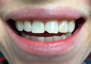
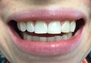
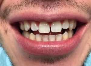
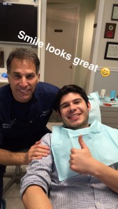
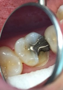
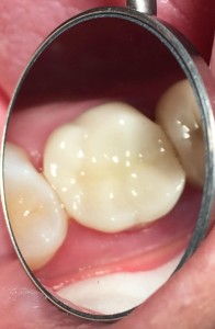
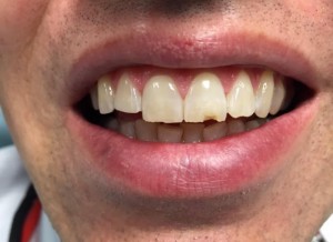
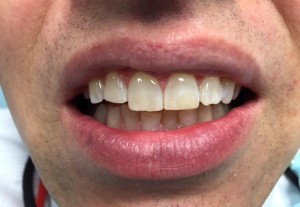
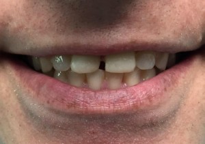
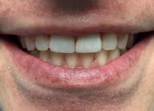
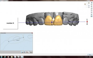
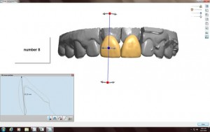
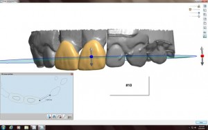
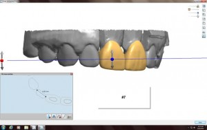
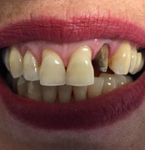
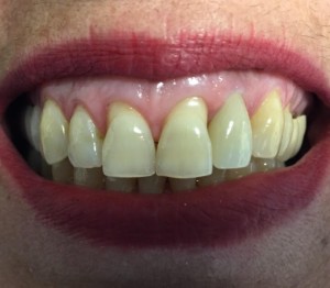
This patient today chipped her front tooth biting a coffee stirrer straw. I was able to restore the tooth with a bonded composite restoration in 30 minutes. Looks great!!!
Tag Archives: arlington
Dr. Gentry’s article in The Elm, The University of Maryland
From The Horse’s Mouth
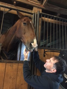
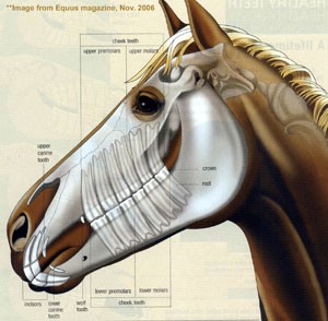
Horses have 2 sets of teeth, 24 baby teeth, which are replaced by 5 years of age by between 36-40 permanent teeth. These consist of 6 upper and 6 lower incisors, 12 upper and 12 lower back premolar and molar teeth. Most male horses also have canine teeth, called fighting teeth, while females do not. There is an interdental space with no teeth, just gums, between the incisors and premolars. This is where the bit part of the bridle is placed.
Horses eat pretty much constantly for 10-12 hours a day and their teeth continuously erupt to offset the wear from the repeated chewing. Young adult horse teeth can be 5 inches long. Most of the tooth is under the gums and it slowly erupts about 1/8 of an inch each year until the horse gets old and eventually the teeth become very short and eventually fall out.
It is possible to tell the approximate age of a horse by its teeth. The expression “Don’t look a gift horse in the mouth” means that it is rude and ungrateful to check the teeth of a horse given to you as a present, as you would one you were considering purchasing. So don’t be ungrateful when you receive a gift.
Horses teeth wear unevenly and develop sharp edges, which irritate and lacerate the tongue and cheeks and make it difficult and painful to eat and have the bit in their mouth. A procedure called floating the horse’s teeth, where an equine dentist smoothes the sharp edges is done 1-2 times a year.
Friday afternoon chipped and broken teeth.
Thursday morning chipped tooth.
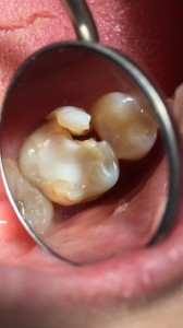
Fractured teeth of the day. Every day I see broken teeth. Here are 2 examples from today.
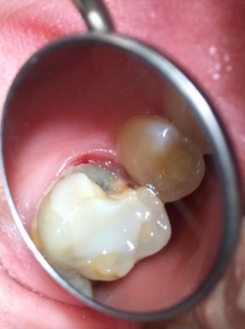
This patient cracked his molar biting a chicken bone. I was able to save the tooth, and without a root canal, but it did require a crown lengthening periodontal surgery and a crown.
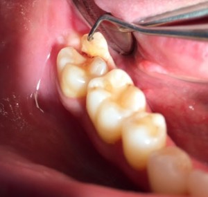
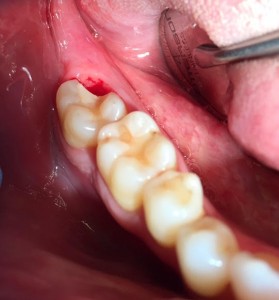
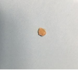
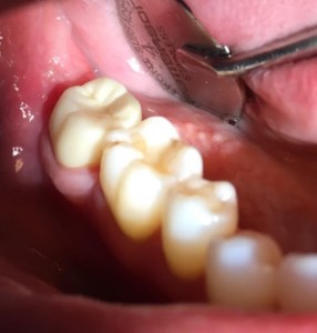
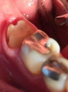
This next patient bit down on a mint candy and fractured her molar down the root. The tooth could not be restored and was extracted and we will place a dental implant.
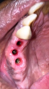
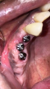
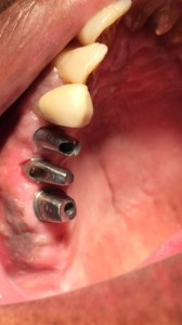
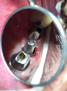
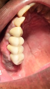
Implant case of the week.
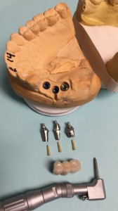
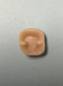
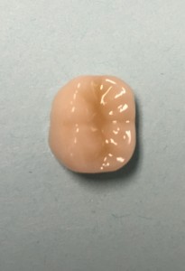
This was a challenging implant restoration case due to the alignment of implants. In order for the implant to osseointegrate firmly into the jaw bone we like to place the implants where the jaw bone is the thickest and strongest. In this case the best bone was not lined up nicely so we had to stagger the implants. I was still able to make the final implant crowns line up perfectly in the mouth.
Before and After
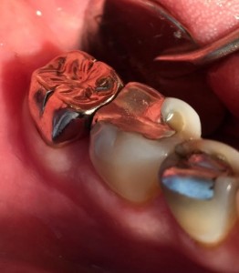
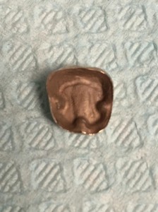
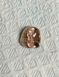
Retention Grooves to help crowns last longer.
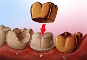
On these 2 crowns, the first porcelain and second gold, that I did last week in my office I show how I place retention grooves in my crown preparations to help lock my crowns in. These grooves prevent rotation and help secure the crowns in place. These slots allows for more surface area for the cement and results in a longer lasting restoration that holds firmly in place. This is especially important in short weak teeth. With this technique I am able to save teeth that otherwise would need to be extracted or require periodontal crown lengthening surgery.
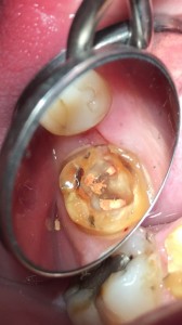
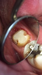
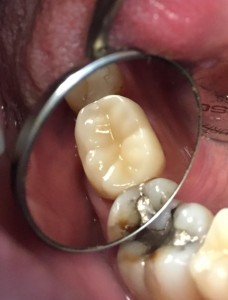
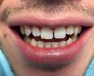
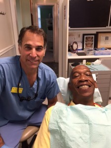
Two patient’s smiles I made happy today.
Cone Beam Computerized Tomography Dental Imaging
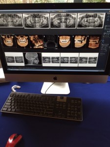
CBCT systems used by dental professionals rotate around the patient, capturing data using a cone-shaped X-ray beam. This data is used to reconstruct a three-dimensional (3D) image of the of the patient’s head and neck. CBCT uses more radiation than regular dental teeth x-rays, but still less than 10% of the radiation used in conventional medical CT scan of the same area.
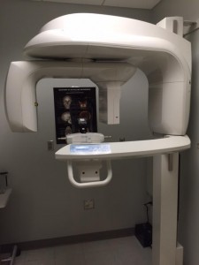
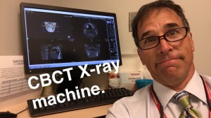
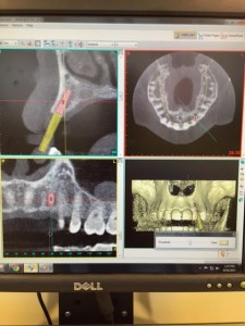
This is the Carestream 9300 Imaging machine we have at the University of Maryland Dental School. I mostly use this to plan dental implant placement.
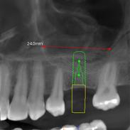
Dentists use Cone Beam CT imaging for the following:• 3-D observation of overall oral/facial bony characteristics, allowing easier diagnosis and placement of dental implants • Surgical guide fabrication for implant placement • 3-D observation of teeth for endodontic diagnosis and treatment • Diagnosis and treatment of tooth impactions • Identification of inferior alveolar nerve and mental foramen location • Identification of the location of the maxillary sinus • Identification of the presence of odontogenic lesions • Trauma evaluation and treatment • Analysis of temporomandibular joint characteristics leading to diagnosis and treatment • Integration with CAD/CAM devices for fabrication of prosthodontics or orthodontic appliances • Identification for referral of numerous conditions or diseases not normally within the realm of dentistry, but that can be shown on typical cone beam images.
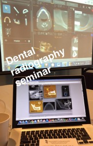
These last 2 pictures show how we use information obtained from the Cone Beam CT to plan placement of dental implants.
