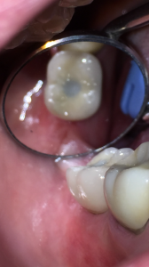
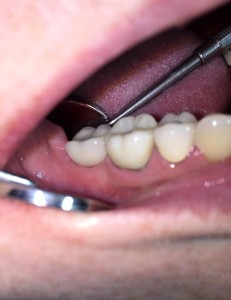
Tag Archives: dentist

Root canals explained.

What is a root canal?

patientendo1

To understand root canal treatment, it helps to know something about the anatomy of the tooth. Inside the tooth, under the white enamel and a hard layer called the dentin, is a soft tissue called the pulp. The pulp contains blood vessels, nerves, and connective tissue and creates the surrounding hard tissues of the tooth during development.

The pulp extends from the crown of the tooth to the tip of the roots where it connects to the tissues surrounding the root. The pulp is important during a tooth’s growth and development. However, once a tooth is fully mature it can survive without the pulp, because the tooth continues to be nourished by the tissues surrounding it

Why would I need a root canal?

A root canal is necessary when the pulp, the soft tissue inside the root canal, becomes inflamed or infected. The inflammation or infection can have a variety of causes: deep decay, or a crack or chip in the tooth. In addition, an injury to a tooth may cause pulp damage even if the tooth has no visible chips or cracks. If pulp inflammation or infection is left untreated, it can cause pain or lead to an abscess.

patientendo2

What are the signs of needing root canal treatment?

Signs to look for include pain, prolonged sensitivity to heat or cold, tenderness to touch and chewing, discoloration of the tooth, and swelling, drainage and tenderness in the lymph nodes as well as nearby bone and gum tissues. Sometimes, however, there are no symptoms.

How does root canal treatment save the tooth?

The dentist removes the inflamed or infected pulp, carefully cleans and shapes the inside of the root canal, then fills and seals the space. Afterwards, a crown or other restoration on the tooth to protect and restore it to full function. After restoration, the tooth continues to function like any other tooth.

Will I feel pain during or after the procedure?

Many root canal procedures are performed to relieve the pain of toothaches caused by pulp inflammation or infection. With modern techniques and anesthetics, most patients report that they are comfortable during the procedure.

For the first few days after treatment, your tooth may feel sensitive, especially if there was pain or infection before the procedure. This discomfort can be relieved with over-the-counter or prescription medications. Your tooth may continue to feel slightly different from your other teeth for some time after your root canal treatment is completed.

Step-by-Step Root Canal Procedure

Root canal treatment can often be performed in one or two visits and involves the following steps:

patientendo3

1. The dentist examines and x-rays the tooth, then administers local anesthetic. After the tooth is numb, a small protective sheet called a “dental dam” is placed over the area to isolate the tooth and keep it clean and free of saliva during the procedure.

patientendo4

2. An opening in the crown of the tooth. Very small instruments are used to clean the pulp from the pulp chamber and root canals and to shape the space for filling.

patientendo5

3. After the space is cleaned and shaped, the dentist fills the root canals with a biocompatible material, usually a rubber-like material called gutta-percha. The gutta-percha is placed with an adhesive cement to ensure complete sealing of the root canals. In most cases, a temporary filling is placed to close the opening. The temporary filling will be removed usually 1 week later.

patientendo6

Can all teeth be saved with a root canal?

Most teeth can be saved. Occasionally, a tooth can’t be saved because the root canals are not accessible, the root is severely fractured, the tooth doesn’t have adequate bone support, or the tooth cannot be restored.

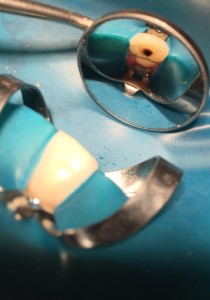
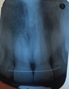
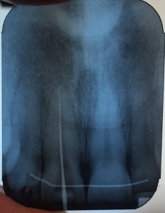
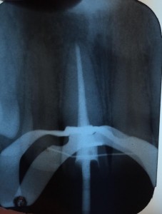
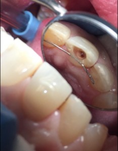
Here’s photos of what an actual root canal looks like on a patient.
unnamed-1
Access opening made by Dr. Gentry on inside surface of tooth.
unnamed-3
Isolation dam placed over tooth.
unnamed-4
Before treatment x-ray.
unnamed-6
Cleaning the root canal.
unnamed-7
Final root canal fill.


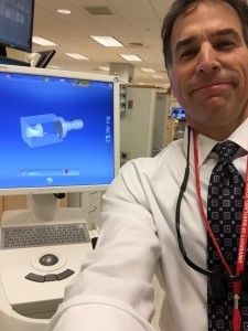
Dr. Gentry explains CAD/CAM technology and 3D printing in dentistry.

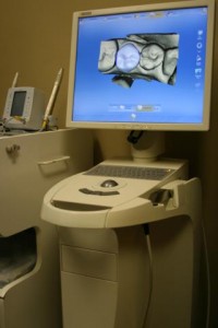
CAD/CAM is an acronym for computer-aided design/computer-aided manufacturing. CAD/CAM technology has been increasingly incorporated into dentistry over the past 20 years. CAD/CAM technology is used by dentists and dental laboratories to provide our patients with 3-D milled ceramic crowns and other types of restorations, and to fabricate abutments for dental implants, used to replace missing teeth. A digital impression is taken of the patient’s teeth or dental impression, and the restoration is milled from a single block of tooth colored ceramic material in a milling chamber.

IMG_1509
Digital design of the crown restoration.
unnamed
Sending the digital impression to the 3-D milling machine
milling-machine
3-D printer fabricating the dental restoration.

The 3-D milling machine fabricating the dental restoration.

teeth whitening-take home trays

opalescence-go_little-miss-fearless_teeth-whitening

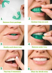
For home teeth whitening, Dr. Gentry recommends Opalescence Go.

Professional results—to go

Opalescence Go is the professional alternative to over-the-counter whitening options that Dr. Gentry recommends, and has for sale in his office for only $50 per kit. It’s a simple, fast, and great-tasting way to start a whitening treatment, or for whitening touch-ups. With no impressions or custom trays necessary, Opalescence Go is ready to use right out of the package!

  • Opalescence tooth whitening gel contains PF (potassium nitrate and fluoride). Potassium nitrate has been shown to help reduce sensitivity. Fluoride has been shown to help reduce cavities and strengthen enamel. Together they help to improve the overall health of the teeth.
  • No impressions, no models, no lab time
  • Sleek and comfortable; adapts to any smile
  • Discreet, clear tray material
  • 10% – wear 30-60 minutes
  • 15% – wear 15-20 minutes
  • Peach, Melon, and Mint flavors


$T2eC16JHJF0FFZpt7V-uBRrkstmle!--60_1

maxresdefault

Invisalign

invisalign5

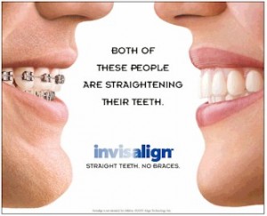
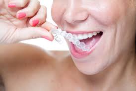
Invisalign® straightens your teeth without wires and brackets, using a series of clear, customized, removable appliances called aligners. It’s virtually undetectable, which means hardly anyone will know that you’re straightening your teeth.

CLEARLY DIFFERENT

The Invisalign System combines advanced 3-D computer graphics technology with 100-year-old science of orthodontics. Invisalign aligners are designed to move your teeth in small steps to the desired final position. Each aligner is precisely calibrated and manufactured to fit your mouth at each stage of the treatment plan. Your first step is to visit our office and see Dr. Jeanette Coutin-Gentry to determine if Invisalign is right for you. Dr. Coutin is an Invisalign Certified Preferred Provider with many years of experience with Invisalign.

After sending precise treatment instructions, Invisalign uses advanced computer Invisalign technology to translate these instructions in a sequence of finely calibrated aligners — as few as 12 or as many as 48. Each aligner is worn for about two weeks and only taken out to eat, brush and floss. As you replace each aligner with the next, your teeth will begin to move gradually — week-by-week until the final alignment prescribed is attained. Then you’ll be smiling like you never have before!

While the results may appear the same—a confident, beautiful smile—when you stop and actually compare Invisalign® to other teeth-straightening options, the advantages become quite apparent. Knowing the pros and cons of each option ahead of time will help you make a more confident decision.
invisalign1

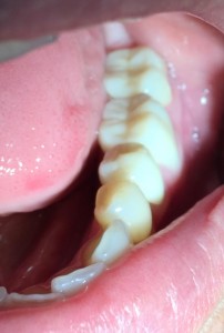
-Invisalign Advantages over Braces –

COMPARISON CHART INVISALIGN/  BRACES
Effectively treats a wide variety of cases, including crowding, spacing, crossbite, overbite and underbite.
Straightens your teeth.
Allows you to eat whatever foods you enjoy.
Lets you remove the device when you want.
Lets you enjoy virtually invisible teeth-straightening.
Allows you to brush and floss your teeth normally for better periodontal health.
Consists of smooth, comfortable plastic instead of sharp metal that is more likely to irritate your cheeks and gums.
Frees up your busy schedule, with office visits only every four to six weeks.
Invisalign Teen®: Provides up to six free replacement aligners if lost or broken.*

images-2

https://youtu.be/m8cMGvWAPZY

Crowns

Dr Gentry cementing some of his porcelain crowns

unnamed-1
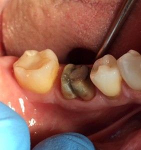
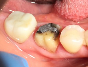
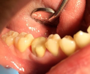
Broken down tooth that had a root canal prepared for crown.
unnamed-3
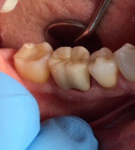
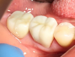
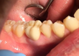
Porcelain crown placed.
unnamed-1
Molar crown prep
unnamed-3
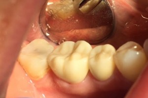
Porcelain crown cemented
unnamed-1
Two teeth prepped for crowns.
unnamed-3
Porcelain crowns cemented.
unnamed-3
Crown preparation
unnamed-1
Crown cemented
unnamed-11
Crown prep
unnamed-12
Crown cemented

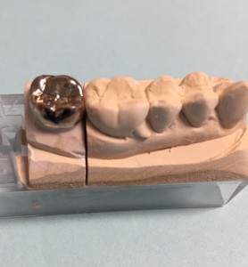
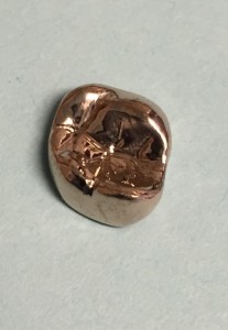
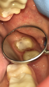
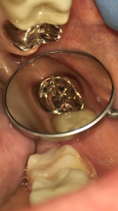
Occasionally Dr. Gentry will recommend a gold crown for a back molar if the patient constantly clenches their teeth or is a severe grinder.

unnamed-3
Gold crown on patient’s stone model.
unnamed-1
The gold crown.
unnamed
The back molar tooth.
unnamed-1
Dr. Gentry cements the gold crown cemented into place.


Cracked teeth/tooth fractures.

unnamed-2
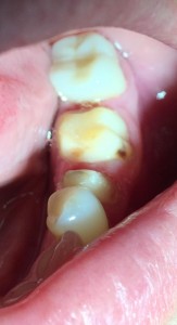
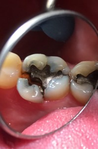
Cracks on back molars.

If when you bite down, you feel a sharp pain you may have a cracked tooth. A tooth crack may have been caused by:

  • Chewing on a hard object or food such as ice, nuts, candy, or popcorn.
  • An accident, like a blow to the mouth.
  • Grinding or clenching your teeth.
  • Weak tooth caused by wear or large old fillings, or root canal.

If you feel pain when you bite down on a tooth please let Dr. Gentry know and he will evaluate the tooth and discuss the best treatment protocol for you. If treated early, the cracked tooth possibly can be treated with a bonded filling or a crown. If the crack is deeper the tooth may require a root canal or even extraction.

unnamed-6
Cracked molar. Teeth weak due to large old fillings and tooth broke biting down on an almond.
unnamed-7
Dr. Gentry was able to save the tooth with a large bonded composite restoration. A crown will be placed on top.
unnamed-6
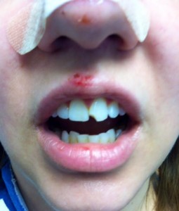
Fractured front tooth caused by an accident.
unnamed-7
Emergency tooth bonding to temporarily repair the fractured front tooth.
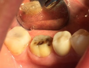
If the crack is deep or extends down the root, the tooth may need to be extracted.

unnamed-8
Cracked tooth caused from grinding teeth.
unnamed-9
Abscess
unnamed-10
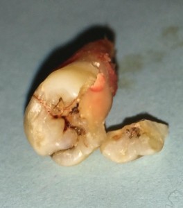
Vertical root fracture. Dr. Gentry had to extract the tooth.
unnamed-1
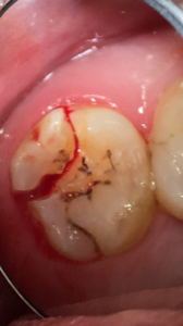
Cracked back molar.
unnamed-11
Crack extended under the gums along root. Dr. Gentry had to extract the molar.

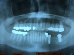
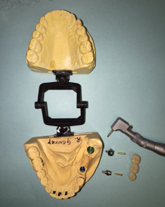
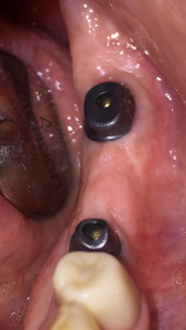
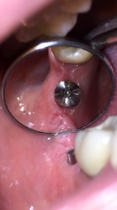
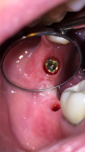
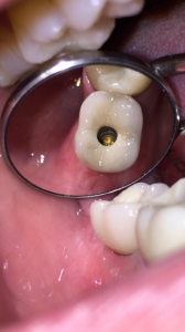
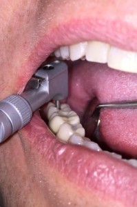
Dr. Gentry demonstrates the steps involved in the restoration of lower second molar Biomet 3i Encode Implant.

unnamed-1
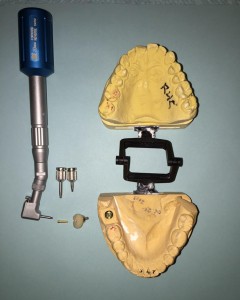
Stone model of the patient’s teeth and implant parts.
unnamed-3
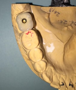
Implant try-in on stone model.
unnamed
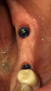
Healing abutment in the mouth.
unnamed-1
Following removal of the healing abutment the implant the implant is visible.
unnamed-2
The implant crown placed.
unnamed-4
Implant crown screw tightened into place.
unnamed-3
Screw access hole filled in.
unnamed-6
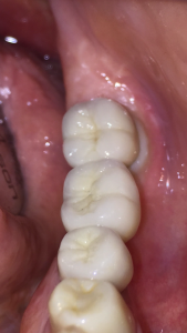
Beautiful implant crown.