CBCT systems used by dental professionals rotate around the patient, capturing data using a cone-shaped X-ray beam. This data is used to reconstruct a three-dimensional (3D) image of the of the patient’s head and neck. CBCT uses more radiation than regular dental teeth x-rays, but still less than 10% of the radiation used in conventional medical CT scan of the same area.
This is the Carestream 9300 Imaging machine we have at the University of Maryland Dental School. I mostly use this to plan dental implant placement.
Dentists use Cone Beam CT imaging for the following:• 3-D observation of overall oral/facial bony characteristics, allowing easier diagnosis and placement of dental implants • Surgical guide fabrication for implant placement • 3-D observation of teeth for endodontic diagnosis and treatment • Diagnosis and treatment of tooth impactions • Identification of inferior alveolar nerve and mental foramen location • Identification of the location of the maxillary sinus • Identification of the presence of odontogenic lesions • Trauma evaluation and treatment • Analysis of temporomandibular joint characteristics leading to diagnosis and treatment • Integration with CAD/CAM devices for fabrication of prosthodontics or orthodontic appliances • Identification for referral of numerous conditions or diseases not normally within the realm of dentistry, but that can be shown on typical cone beam images.
These last 2 pictures show how we use information obtained from the Cone Beam CT to plan placement of dental implants.
It is true that sugar does cause cavities, but it’s not the sugar directly. The naturally occurring streptococcus bacteria that live in our mouths consume the sugar, ferment it, and produce acid such as lactic acid. It is these acids that cause teeth demineralization and the formation of cavities. So it is actually the acid from the bacteria in plaque in our mouth that eats the holes in our teeth causing cavities.
Here’s the interesting part, it’s not the total amount of sugar we eat, but the total amount of time the sugar is in contact with our teeth. It is preferable to have a piece of pumpkin pie with a scoop of ice cream for Thanksgiving dessert, than sip on a soda all afternoon. It is better to have a few sugar cookies, or slice of Christmas apple pie, than to repeatedly sip coffee with sugar all morning long. One Altoids mint or cough drop per hour throughout the day is ten times more cavity producing than 1 big piece of cake for dessert, even though the cake has much more sugar and calories.
The repeated cycles of eating sugar and acid formation is what is the key. It is the frequency, or the amount of time the sugar is in the mouth, not the total amount of sugar. So do enjoy a few scrumptious (and quick) holiday desserts, just please make sure to brush and floss after every meal and visit your dentist regularly. Happy Holidays!!!
Philip A. Gentry, DDS, FAGD

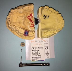
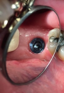
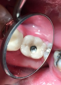
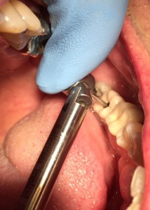
In this case report Dr. Gentry demonstrates the importance of taking a radiograph when seating the custom abutment and crown before final torquing of the gold implant interface connecting screw.
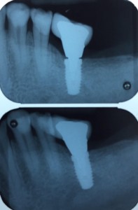
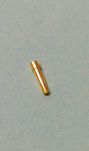
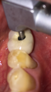
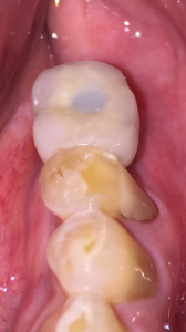
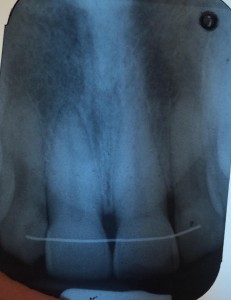
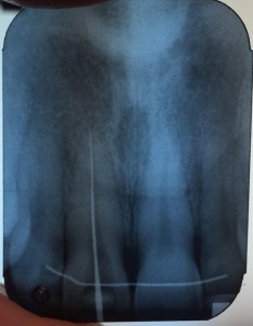
Here’s an example from a case I was doing with a dental student involving the 2 upper front teeth. In the first x-ray the impression copings were not fully seated. I re-positioned the copings and another x-ray was taken to confirm proper alignment. You cannot tell without an x-ray since this is below the gum-line.
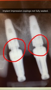
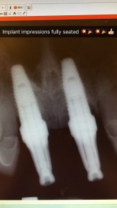
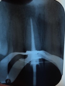
Here’s an example of another implant check x-ray to verify proper seating.

To understand root canal treatment, it helps to know something about the anatomy of the tooth. Inside the tooth, under the white enamel and a hard layer called the dentin, is a soft tissue called the pulp. The pulp contains blood vessels, nerves, and connective tissue and creates the surrounding hard tissues of the tooth during development.
The pulp extends from the crown of the tooth to the tip of the roots where it connects to the tissues surrounding the root. The pulp is important during a tooth’s growth and development. However, once a tooth is fully mature it can survive without the pulp, because the tooth continues to be nourished by the tissues surrounding it
A root canal is necessary when the pulp, the soft tissue inside the root canal, becomes inflamed or infected. The inflammation or infection can have a variety of causes: deep decay, or a crack or chip in the tooth. In addition, an injury to a tooth may cause pulp damage even if the tooth has no visible chips or cracks. If pulp inflammation or infection is left untreated, it can cause pain or lead to an abscess.

Signs to look for include pain, prolonged sensitivity to heat or cold, tenderness to touch and chewing, discoloration of the tooth, and swelling, drainage and tenderness in the lymph nodes as well as nearby bone and gum tissues. Sometimes, however, there are no symptoms.
The dentist removes the inflamed or infected pulp, carefully cleans and shapes the inside of the root canal, then fills and seals the space. Afterwards, a crown or other restoration on the tooth to protect and restore it to full function. After restoration, the tooth continues to function like any other tooth.
Many root canal procedures are performed to relieve the pain of toothaches caused by pulp inflammation or infection. With modern techniques and anesthetics, most patients report that they are comfortable during the procedure.
For the first few days after treatment, your tooth may feel sensitive, especially if there was pain or infection before the procedure. This discomfort can be relieved with over-the-counter or prescription medications. Your tooth may continue to feel slightly different from your other teeth for some time after your root canal treatment is completed.
Root canal treatment can often be performed in one or two visits and involves the following steps:

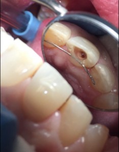
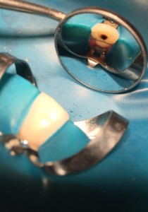
1. The dentist examines and x-rays the tooth, then administers local anesthetic. After the tooth is numb, a small protective sheet called a “dental dam” is placed over the area to isolate the tooth and keep it clean and free of saliva during the procedure.

2. An opening in the crown of the tooth. Very small instruments are used to clean the pulp from the pulp chamber and root canals and to shape the space for filling.

3. After the space is cleaned and shaped, the dentist fills the root canals with a biocompatible material, usually a rubber-like material called gutta-percha. The gutta-percha is placed with an adhesive cement to ensure complete sealing of the root canals. In most cases, a temporary filling is placed to close the opening. The temporary filling will be removed usually 1 week later.

Most teeth can be saved. Occasionally, a tooth can’t be saved because the root canals are not accessible, the root is severely fractured, the tooth doesn’t have adequate bone support, or the tooth cannot be restored.

Invisalign® straightens your teeth without wires and brackets, using a series of clear, customized, removable appliances called aligners. It’s virtually undetectable, which means hardly anyone will know that you’re straightening your teeth.
The Invisalign System combines advanced 3-D computer graphics technology with 100-year-old science of orthodontics. Invisalign aligners are designed to move your teeth in small steps to the desired final position. Each aligner is precisely calibrated and manufactured to fit your mouth at each stage of the treatment plan. Your first step is to visit our office and see Dr. Jeanette Coutin-Gentry to determine if Invisalign is right for you. Dr. Coutin is an Invisalign Certified Preferred Provider with many years of experience with Invisalign.
After sending precise treatment instructions, Invisalign uses advanced computer Invisalign technology to translate these instructions in a sequence of finely calibrated aligners — as few as 12 or as many as 48. Each aligner is worn for about two weeks and only taken out to eat, brush and floss. As you replace each aligner with the next, your teeth will begin to move gradually — week-by-week until the final alignment prescribed is attained. Then you’ll be smiling like you never have before!
While the results may appear the same—a confident, beautiful smile—when you stop and actually compare Invisalign® to other teeth-straightening options, the advantages become quite apparent. Knowing the pros and cons of each option ahead of time will help you make a more confident decision.
-Invisalign Advantages over Braces –
| COMPARISON CHART | INVISALIGN/ | BRACES |
|---|---|---|
| Effectively treats a wide variety of cases, including crowding, spacing, crossbite, overbite and underbite. |  |
 |
| Straightens your teeth. |  |
 |
| Allows you to eat whatever foods you enjoy. |  |
|
| Lets you remove the device when you want. |  |
|
| Lets you enjoy virtually invisible teeth-straightening. |  |
|
| Allows you to brush and floss your teeth normally for better periodontal health. |  |
|
| Consists of smooth, comfortable plastic instead of sharp metal that is more likely to irritate your cheeks and gums. |  |
|
| Frees up your busy schedule, with office visits only every four to six weeks. |  |
|
| Invisalign Teen®: Provides up to six free replacement aligners if lost or broken.* |  |
If you or someone you know experiences severe tooth, jaw or facial muscle pain, it may be from the effects of grinding or clenching your teeth. The good news is that Dr. Gentry may be able to prescribe a custom-made Comfort H/S™ (Hard/Soft) Bite Splint, which offsets the pain caused by bruxing or grinding your teeth.
A nightguard is a proactive step to protect your existing healthy teeth. A clear, thin removable device, your custom-made bite splint is worn over your lower or upper teeth as you sleep.
Studies suggest those who grind and clench their teeth may experience up to 80 times the normal tooth wear per day compared to those who do not. The good news is that a simple bite splint can offset the effects of this often-subconscious habit while protecting your teeth from daily wear and tear.